Optomap Retinal Imaging
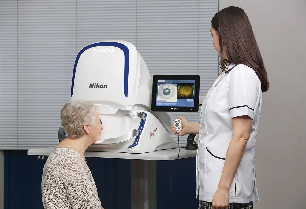
A single optomap scan captures up to 200° of your retina — the wide periphery a standard view can miss — in just seconds, and often without dilating drops. We use the Optos California to screen for and document retinal health at our Newton, MA office, serving Needham, Wellesley, Brookline, Waltham, and Greater Boston.
of your retina in a single optomap capture
screening add-on to a comprehensive exam
color + autofluorescence (optomap af) imaging
What is optomap?
The retina is the light-sensitive tissue lining the back of your eye. A traditional view through the pupil shows only the central portion of it at a time — the far edges are harder to see. Optomap is an ultra-widefield scan, taken with the Optos California, that captures up to 200 degrees of the retina in a single digital image.
That matters because many important findings — retinal tears, early diabetic changes, thinning, and lesions — first appear in the periphery. Optomap makes that region quick to see and, just as importantly, easy to save and compare from year to year so subtle changes stand out.
Alongside standard color imaging, the Optos California also captures autofluorescence (optomap af), which reveals the health of the retinal pigment cells and can highlight disease that isn't obvious on a color photo alone.
See more of your retina
One capture takes a fraction of a second per eye and records the optic nerve, macula, and the wide periphery together — a documented baseline you and Dr. Patel can compare year over year.
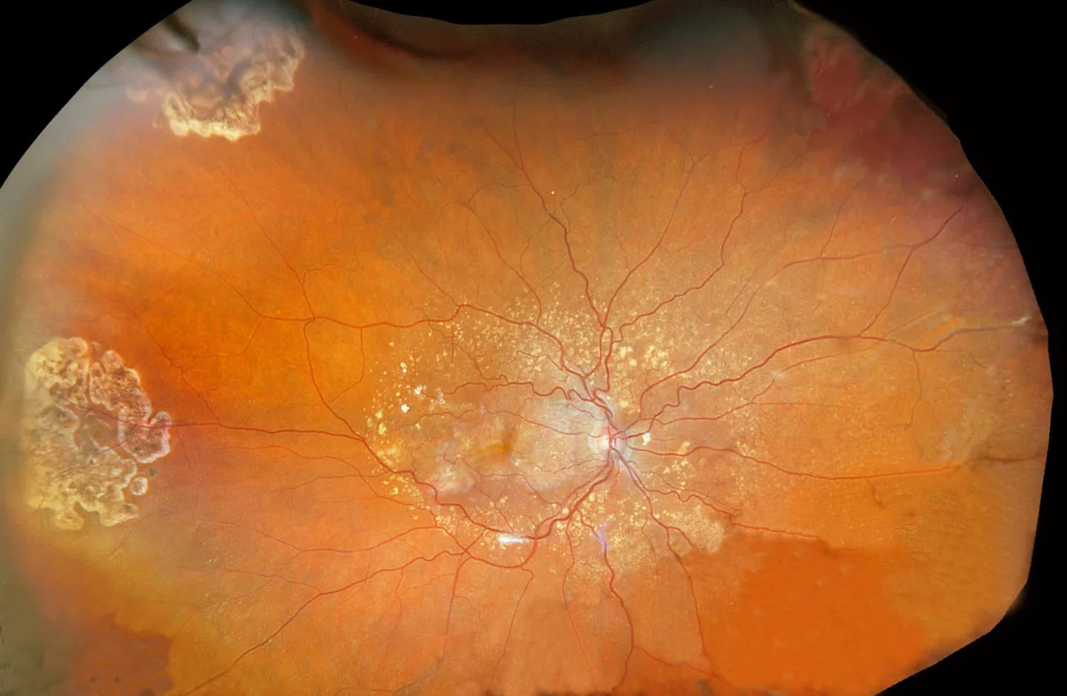
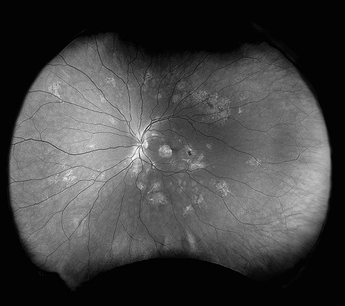
Optomap images courtesy of Optos, shown as examples of ultra-widefield imaging.
What optomap can help reveal
Imaging supports the doctor's exam in screening for a range of eye and systemic conditions.
Diabetic retinopathy
Early bleeding and vascular changes from diabetes, including in the periphery.
Retinal tears & detachment
Peripheral tears, holes, and thinning that can precede a sight-threatening detachment.
Macular degeneration
Changes at the macula that affect central, detailed vision — tracked over time.
Hypertensive changes
Signs of high blood pressure and other systemic conditions visible in the retinal vessels.
Retinal nevi & melanoma
Freckles and pigmented lesions of the retina that need documentation and monitoring.
Optic nerve & more
A documented view of the optic nerve to support glaucoma and overall eye-health assessment.
Imaging is a screening and documentation tool that supports — but does not replace — a doctor's clinical examination and diagnosis.
Optomap and a dilated exam
A common question is whether optomap replaces having your eyes dilated. The honest answer: they do different things, and they're strongest together.
The optomap scan
- Wide, 200° documented image in seconds
- Often no dilating drops needed
- Easy to save and compare year to year
- Excellent for screening and monitoring
A dilated exam
- Lets the doctor view the retina in 3-D, live
- Direct inspection of the far periphery
- Preferred when a closer look is warranted
- Recommended for certain symptoms and risks
Bottom line: optomap complements a dilated exam — it doesn't universally replace it. For many patients it means a thorough, documented look at the retina without drops. When your eyes call for dilation, Dr. Patel will still recommend it.
If you have diabetes, we do both
Diabetes can quietly damage the retina, and some of the earliest changes appear in the periphery. That's why, for our patients with diabetes, we perform both a dilated exam and optomap ultra-widefield imaging at every visit — each catches things the other can miss.
Research using ultra-widefield imaging finds significantly more diabetic retinopathy lesions than standard photography, and lesions concentrated in the peripheral retina are linked to a higher risk of the disease progressing.1 Seeing and documenting that periphery helps us catch trouble sooner and monitor it closely.
Dilated exam
A direct, three-dimensional look at the retina at every diabetic visit.
Optomap imaging
A wide, documented capture of the periphery to catch and track change.
Simple, Transparent Pricing
Add optomap ultra-widefield imaging to your comprehensive eye exam. An optional screening enhancement — no surprise fees.
Book Your ExamCommon Questions
What patients ask most about optomap imaging.
I just want a regular eye exam — do I really need retinal imaging or dilation?
Checking the health of your retina is part of a regular eye exam — not an extra. A complete exam does two jobs: it measures how well you see, and it checks whether your eyes are healthy. The retina is where the most serious eye problems — retinal tears, diabetic changes, glaucoma damage, even melanoma — first appear, often with no symptoms and no change in your vision until late. There are two ways to examine it: dilating drops or retinal imaging, and sometimes both. An exam that skips both isn't a more “normal” exam — it's an exam with one of its most important parts left out. Optomap simply makes that essential step quick, comfortable, and documented, so we can compare your retina year over year.
What is optomap ultra-widefield retinal imaging?
Optomap is a digital retinal scan taken with the Optos California device that captures up to 200 degrees of your retina in a single image — far more of the back of the eye than a standard view. The scan is non-invasive, takes only seconds per eye, and in many cases doesn't require dilating drops. It includes color imaging and autofluorescence (optomap af), which highlights changes in the retinal cells.
I just want a regular eye exam — do I really need retinal imaging or dilation?
At Newton Advanced Eye Services, optomap ultra-widefield imaging is offered as a $39 screening add-on to a comprehensive eye exam. It's an optional enhancement to your visit. Call (617) 965-2540 with any questions about your specific exam.
Is optomap better than having my eyes dilated?
They serve different purposes and work best together. Optomap captures a wide, documentable image of the retina quickly and often without drops, which is excellent for screening and for tracking changes over time. A dilated exam lets the doctor view the retina in three dimensions and inspect the far periphery directly. Optomap complements a dilated exam — it doesn't universally replace it. When dilation is needed for your eyes, we'll still recommend it.
Do I still need my eyes dilated if I get an optomap?
Often the optomap scan itself doesn't require dilation, which many patients appreciate. Whether you also need a dilated exam depends on your eyes, your symptoms, and your risk factors. If you have diabetes, we perform both a dilated exam and optomap imaging at every visit, because each adds information the other can miss.
Is optomap imaging worth it?
For most people, yes. Optomap lets us see and document far more of the retina in seconds, catch peripheral problems earlier, and compare your images year to year to spot subtle change. It's especially valuable if you have diabetes, high myopia, a family history of retinal disease, or simply want a thorough, documented look at your eye health.
1Ashraf M, Cavallerano JD, Sun JK, Silva PS, Aiello LP. Ultrawide field imaging in diabetic retinopathy: exploring the role of quantitative metrics. J Clin Med. 2021;10(15):3300. doi:10.3390/jcm10153300. This page is educational and does not replace a professional eye examination.
Especially valuable for higher myopia
Higher myopia raises the risk of retinal tears and detachment, which is one reason ultra-widefield imaging pairs so well with myopia care.
A wider view of your eye health
Add optomap to your next comprehensive exam for a fast, comfortable, documented look at your whole retina. Book with Dr. Patel in Newton.